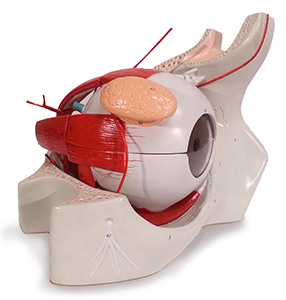
This model is 6 times life-size and dissectible into eight parts. The eyeball sits in its bony socket to provide a better understanding of its relationship with major blood vessels, muscles and nerves. All six muscles of movement can be seen and the upper half of the eyeball with its lacrimal (tear) gland lifts off. Present and extractable are the superior and lateral rectus muscles, the iris and cornea, which can be removed as a unit, the Lucite lens, which actually magnifies and inverts images, and the transparent vitreous body, which exposes the fovea centralis on the retina. The choroid coat, retina, and optic disk can also be seen, as well as a schematic cross section through retinal layers.
Location:
Main Circulation Desk (SRC, Library, 2nd Floor)
Number available:
2