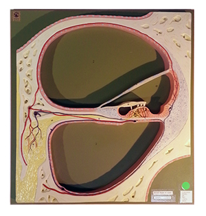
The model depicts a section through the central spiral of the cochlea and has been enlarged approximately 350 times. It shows the vestibular, cochlear, and tympanic ducts; Corti’s organ with hair cells and tectorial and basilar membranes; and spiral ganglia, nerves, and arteries of the ear.
Location:
Main Circulation Desk (SRC, Library, 2nd Floor)
Number available:
2
